Bile Solubility Test Principle, Procedure, Result
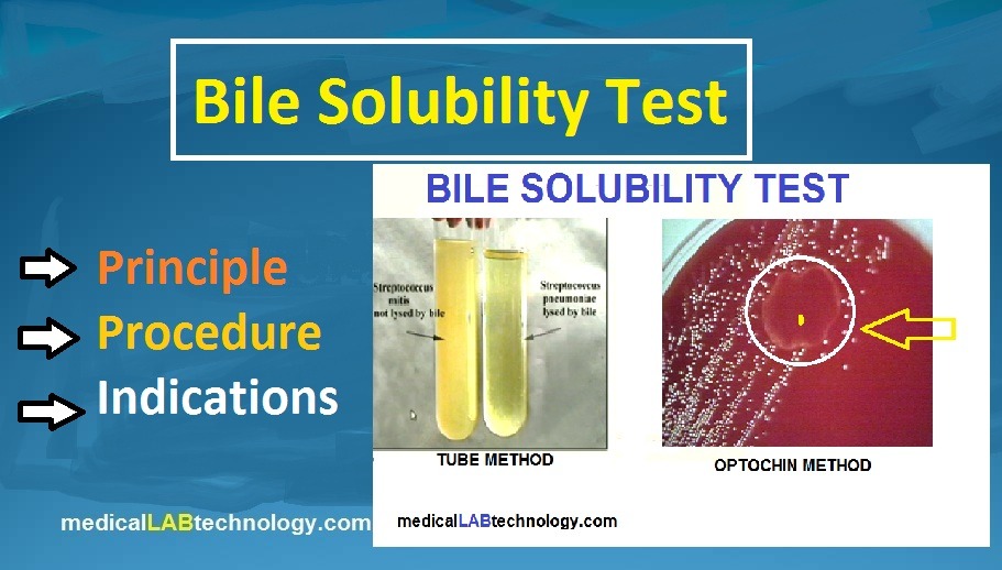
The Bile solubility test is important for the identification of Streptococcus pneumoniae from Viridans types of other streptococci. Principle of bile Solubility test Some bacteria (Strept Pneumoniae) have an autolytic enzyme that autolyzes the broth culture within a few days. while other alpha-hemolytic bacteria are resistant to bile. Bile Solubility test reagents and media The … Read more