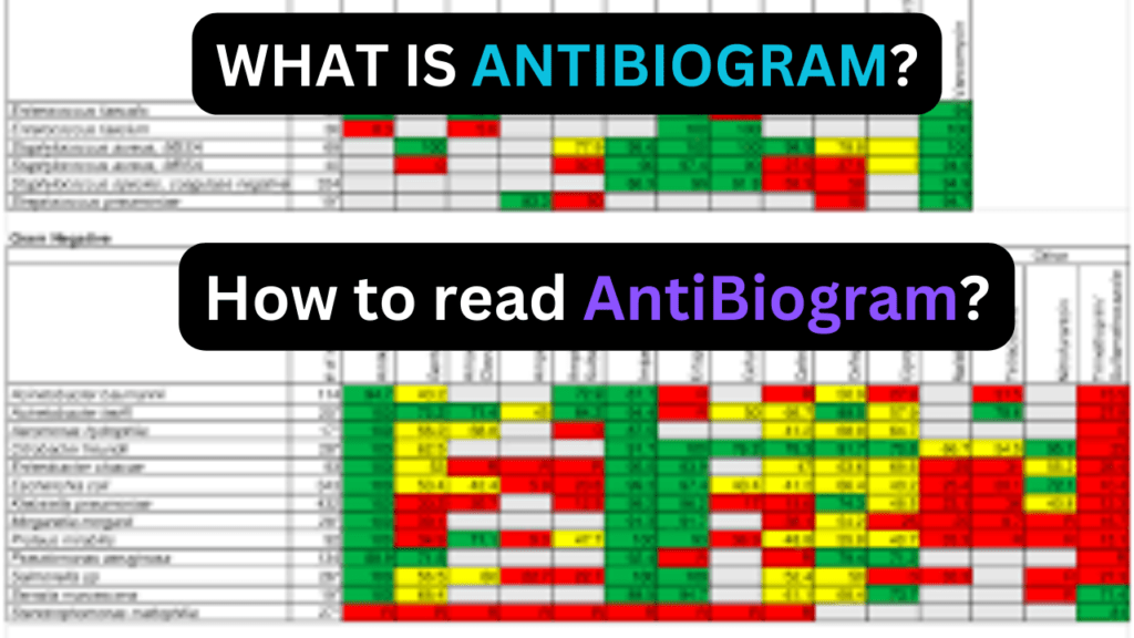
An antibiogram is a laboratory test that helps to choose the most effective antibiotic for treating a bacterial infection. It provides information about the susceptibility of bacteria isolated from a patient to different antibiotics.

Here are the steps to read the antibiogram.
- Bacterial Isolates: The first column typically lists the names or codes of the bacteria that have been isolated from a patient sample. Each row corresponds to a specific bacterium.
- Antibiotics: The top row of the antibiogram lists the names of various antibiotics. These may include common antibiotics such as penicillin, ciprofloxacin, amoxicillin, etc.
- Zone of Inhibition: The body of the antibiogram consists of a grid or table where the intersection of the bacterial isolate and the antibiotic shows the “zone of inhibition. Larger zones usually indicate greater susceptibility to the antibiotic.
Interpretation of the antibiogram result
- Resistant (R): If there is no zone of inhibition or a very small zone, the bacteria are considered resistant to that antibiotic.
- Intermediate (I): An intermediate result suggests that the effectiveness of the antibiotic is uncertain, and it may depend on factors such as the concentration of the drug at the injection site.
- Susceptible (S): A larger zone of inhibition indicates susceptibility, meaning the antibiotic is likely to be effective against the bacteria.
- Minimum Inhibitory Concentration (MIC): MIC may be reported. MIC is the lowest concentration of an antibiotic that inhibits the visible growth of bacteria. Lower MIC values indicate greater sensitivity.
- Clinical Correlation: The final decision on antibiotic choice should consider the clinical context, patient factors, and the severity of the infection.