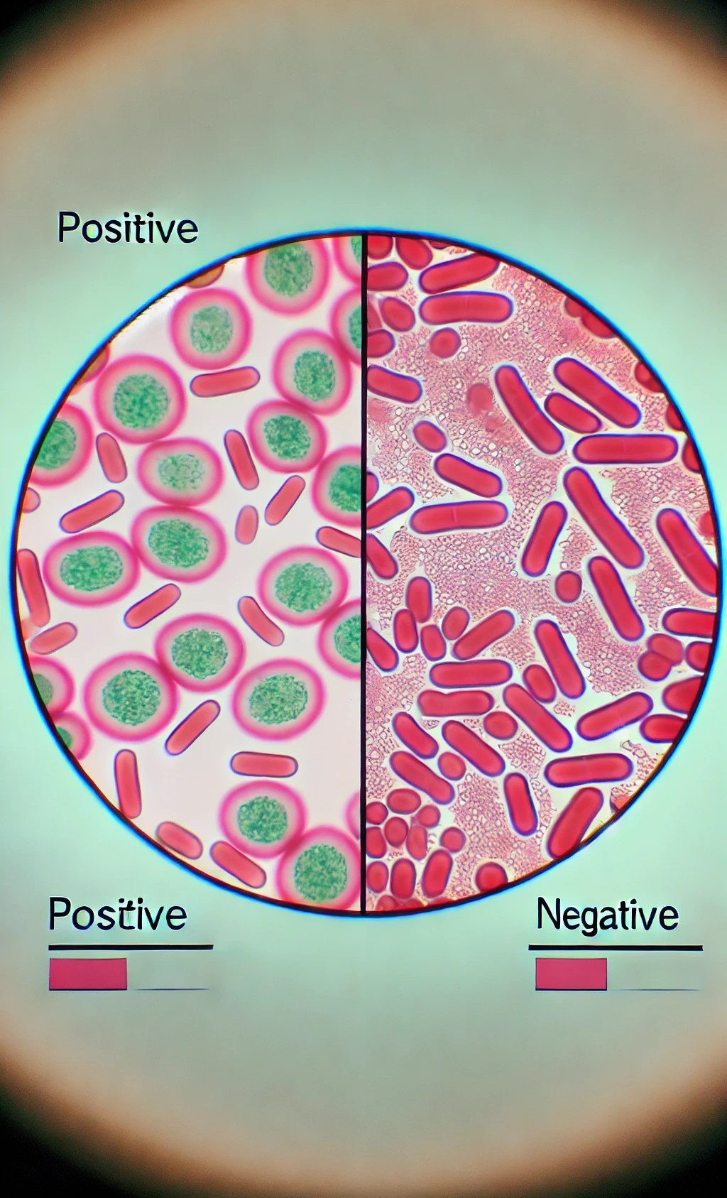
The Endospore Test (or Spore Staining) is a specialized microbiological technique used to detect and visualize bacterial endospores, which are highly resistant structures produced by some bacteria (e.g., Bacillus and Clostridium species).
These endospores protect bacteria from harsh conditions. The most common method for detecting endospores is the Schaeffer-Fulton Method.
Schaeffer-Fulton Endospore Staining Procedure
Materials
- Bacterial culture (on agar or broth).
- Clean glass slides.
- Inoculating loop or needle.
- Heat source (Bunsen burner).
- Malachite green stain.
- Safranin stain (counterstain).
- Distilled water.
- Staining rack.
- Bibulous paper or blotting paper.
Staining Procedure steps
1. Preparation of Smear:
- a. Place a drop of sterile water on a clean glass slide.
- b. Using a sterile inoculating loop, collect a small amount of the bacterial culture and mix it into the water drop to make a thin smear.
- c. Allow the smear to air dry completely.
- d. Heat-fix the smear by passing the slide over the flame of a Bunsen burner several times (about 3–4 times), smear-side up.
2. Primary Staining with Malachite Green:
- Place the slide on a staining rack.
- Cover the smear with malachite green stain.
- Heat the slide gently by passing the Bunsen burner flame under it until steam rises . Keep the slide steaming .
- for 5–10 minutes. Add more malachite green as needed to keep the smear moist.
- Allow the slide to cool for 1–2 minutes.
3. Rinse the slide thoroughly with distilled water to remove excess malachite green. Water will remove the stain from vegetative cells but not from endospores.
4. Counterstaining with Safranin:
- a. Flood the slide with safranin (the counterstain) and let it sit for 30 seconds to 1 minute.
- b. Rinse the slide gently with distilled water to remove excess safranin.
5. Dry and Observe:
- a. Gently blot the slide dry with bibulous paper.
- b. Observe the slide under a light microscope, starting with the lower magnification and moving to oil immersion (1000x) for detailed observation.
Results Interpretation
- Endospores: Appear as small, round, or oval structures and will be green because they retain the malachite green stain.
- Vegetative cells: Appear pink or red due to the safranin counterstain.
