Here are the light microscope vs electron microscope comparisons.
Microscopes have been invaluable tools in the world of science for centuries, enabling researchers to explore the intricate details of the microcosm. Zacharias Janssen (c. 1580): A Dutch spectacle maker, Janssen is often credited with inventing the compound microscope.
While Max Knoll and Ernst Ruska (1931) a German engineer Max Knoll and Ernst Ruska are generally credited with building the first electron microscope. Each type of microscope has its own set of advantages and limitations, making them suitable for different applications.
In this article, we will hunt through the differences between light microscopes and electron microscopes, highlighting their respective strengths and weaknesses.
Light Microscope
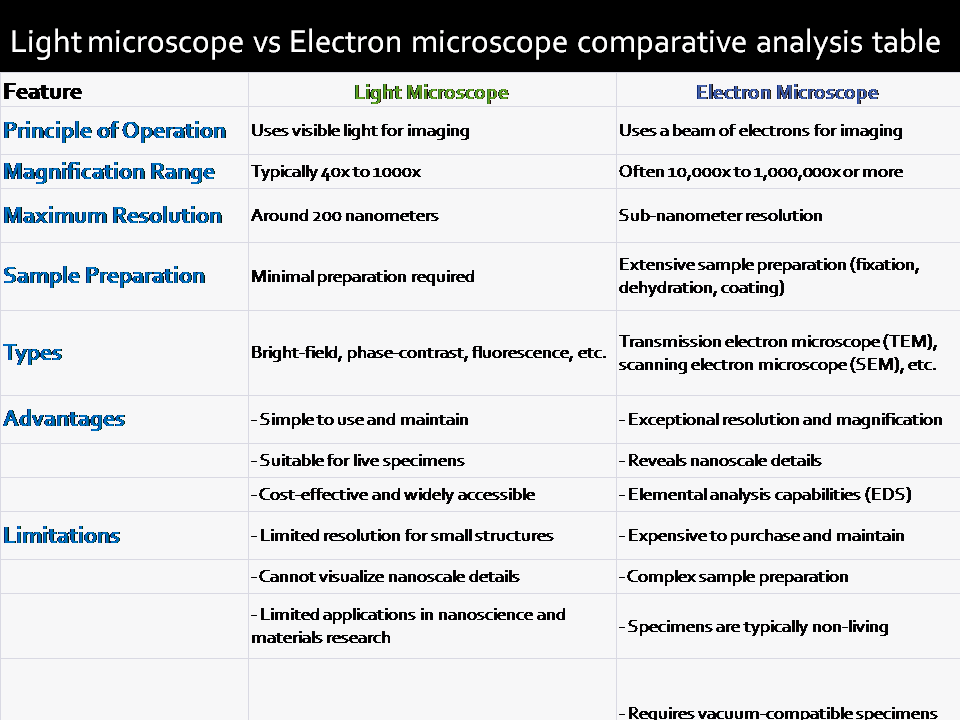
- Principle of Operation:
- Light microscopes, also known as optical microscopes, employ visible light to illuminate and magnify specimens.
- They use lenses to refract light, making the object appear larger to the observer.
- The magnification power of the light microscope
- Light microscopes typically provide magnifications ranging from 40x to 1000x.
- The maximum resolution of light microscopes is around 200 nanometers.
- Sample Preparation:
- Samples for light microscopy require minimal preparation.
- Living specimens can often be observed without extensive processing.
- Types of light microscope
- Common types include bright-field, phase-contrast, and fluorescence microscopes.
- Phase-contrast and fluorescence microscopes enhance contrast and are suitable for specific applications.
- Advantages:
- Relatively simple to use and maintain.
- Suitable for observing living organisms in their natural state.
- Cost-effective and widely accessible.
- Limitations light microscope
- Limited resolution, making it challenging to observe structures smaller than 200 nanometers.
- Cannot provide detailed information about the ultrastructure of cells.
Electron Microscope
- Principle of Operation:
- Electron microscopes use a beam of electrons instead of visible light for imaging.
- Electromagnetic lenses manipulate the electron beam to produce high-resolution images.
- Magnification:
- Electron microscopes offer much higher magnifications, often ranging from 10,000x to 1,000,000x or more.
- They can achieve sub-nanometer resolution, allowing the visualization of ultrafine details.
- Sample Preparation:
- Specimens must undergo extensive preparation, including fixation, dehydration, and embedding in resin.
- Samples are typically coated with a thin layer of metal to enhance electron scattering.
- Types of electron microscope
- Transmission electron microscopes (TEM) and scanning electron microscopes (SEM) are the two main categories.
- TEM is used for thin-section imaging, while SEM provides three-dimensional surface information.
- Advantages of electron microscope
- Exceptional resolution and magnification, allow the study of nanoscale structures.
- Can reveal details about cell organelles, nanoparticles, and material surfaces.
- Wide range of imaging modes, such as energy-dispersive X-ray spectroscopy (EDS) for elemental analysis.
- Limitations of electron microscope
- Expensive to purchase and maintain.
- Complex sample preparation requirements.
- Specimens are typically non-living and must be vacuum-compatible.
Uses of the electron microscope and light microscope
Light microscopes are ideal for biological studies involving live specimens, routine laboratory work, and educational purposes due to their simplicity and affordability. However, their limited resolution makes them less suitable for examining subcellular structures and nanoparticles.
In contrast, electron microscopes are indispensable in fields like nanotechnology, materials science, and cell biology, where high-resolution imaging is crucial. They provide unparalleled insights into the nanoworld but require specialized facilities and skilled operators.
In conclusion, the choice between a light microscope and an electron microscope depends on the specific research goals and available resources. Light microscopes are excellent for everyday applications, while electron microscopes are the go-to tool for investigating the nanoscale world. Together, they form a powerful duo that continues to drive scientific discovery across various disciplines.
3 thoughts on “Light microscope vs Electron microscope comparative analysis table”