Decalcification is a process used to remove calcium salts from tissue samples. This is primarily done in histology and pathology laboratories. It makes the samples suitable for further processing and microscopic examination.
There are many methods, which used to check the decalcification of the different types of tissues. Here we will learn about “End point of decalcification in histopathology”.
Principle of decalcification in histopathology
Decalcified tissue (bone) is placed in the decalcifying agents. If tissue is partially calcified then a white precipitate of calcium appears. But if there is a lack of precipitate then the tissue is calcium-free.
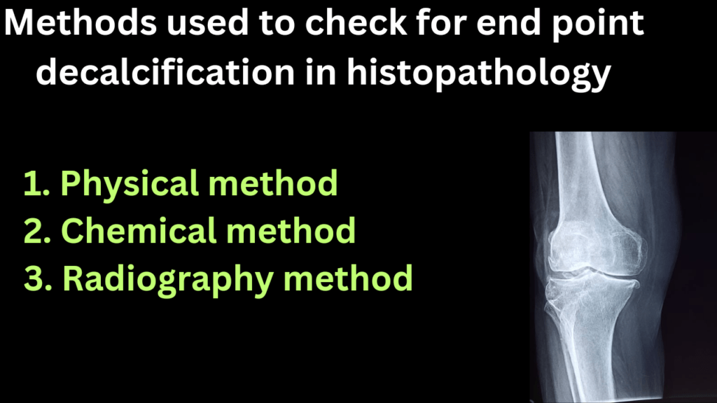
Mention methods to check for end point decalcification in histopathology
There are three methods to assess the end point decalcification.
Note: Physical and x-ray methods are cumbersome, therefore the chemical method is the most commonly used method.
Physical method of decalcification procedure
There are two techniques used in physical method.
- Visual assessment
- Manual Testing
In visual inspection method, sample is visually inspected for any remaining signs of calcification. Fully decalcified tissue will appear soft and pliable, without any visible areas of white or gritty deposits.
On the other hand, we can manually test the tissue’s flexibility. We can also assess its hardness using a fine probe or a pair of forceps. Gently press or poke the tissue to assess its texture.
X-ray method method of decalcification histopathology
X-ray method used to evaluate the extent of calcification in the tissue samples. Take an X-ray image of the tissue prior to decalcification and compare it to the post-decalcification X-ray.
If there are no visible signs of calcification in the post-decalcification image, it suggests successful decalcification.
Chemical method of decalcification
Chemical methods of decalcification involve the use of chemical solutions to remove calcium salts from tissue samples. These techniques are commonly employed in histology pathology laboratories. Here are some common chemical methods of decalcification:
- Acid Decalcification of chemical method
- Chelating Agents for chemical method
Acid decalcification is the most widely used chemical method for decalcification. Tissue samples are immersed in a decalcifying solution containing acids like formic acid, hydrochloric acid, or nitric acid.
The acid reacts with the calcium salts, dissolving them and removing the mineralized components from the tissue.
On the other side, Chelating agents such as ethylene diamine tetraacetic acid (EDTA) can be used for decalcification. These agents form complexes with calcium ions, effectively sequestering them and facilitating their removal from the tissue.
Chelating agents are typically used for decalcifying delicate or smaller tissue teeth. These agents are also used for calcified soft tissues that may be more sensitive to acid-based decalcification.
Importance of decalcification in histopathology
Decalcification plays a crucial role in histopathology for following reasons.
- Tissue Processing: Decalcification is necessary for tissues that contain calcified structures, such as bones, teeth, and calcified soft tissues.It makes the tissues pliable and easier to handle during subsequent processing steps.
- Microscopic Examination: Decalcification enables the visualization of cellular and tissue structures under a microscope. By removing the calcium salts, the tissue becomes transparent and allows for better penetration of stains and dyes.
- It facilitates the identification of cellular details, pathological changes, and the accurate diagnosis of various diseases.
- Special Staining Techniques: Decalcification allows for various special staining techniques in histopathology. These techniques highlight certain tissue components, such as connective tissue, minerals, or pathological features.
- Decalcifing agents removes interfering calcium salts, allowing these stains to bind effectively and reveal additional diagnostic information.
- Molecular Studies: Calcium salts can interfere with nucleic acid extraction and degrade molecular material. Proper decalcification ensures the extraction of high-quality genetic material for molecular investigations.
Method of decalcification in histopathology
There are several methods of decalcification are used to prepare tissue samples for microscopic examination. The choice of method depends on factors like the type of tissue and degree of calcification. Preservation of tissue morphology and downstream applications also play a role. Here are some methods of decalcification used in histopathology:
- Acid Decalcification
- Chelating Agents
- Physical Methods
- Enzymatic Decalcification
Acid Decalcification method
It involves immersing the tissue sample in an acidic solution to dissolve the calcium salts. Commonly used acids include formic acid, hydrochloric acid, and nitric acid. The duration of decalcification varies depending on the size and degree of calcification of the tissue.
Chelating Agents
Ethylene diamine tetra acetic acid (EDTA), can be used for decalcification. These agents form stable complexes with calcium ions, effectively removing them from the tissue. EDTA decalcification is often used for delicate tissues or when preservation of tissue morphology is critical.
Physical Method
Physical methods involve mechanical disruption to break down the calcified tissue. This can include grinding, pulverizing, or drilling the sample to mechanically disrupt and remove the calcium salts.
Enzymatic Decalcification
It uses enzymes, such as trypsin or collagenase, to degrade the organic matrix surrounding the calcium salts. This method selectively removes the organic components, facilitating subsequent decalcification.
Why is it important to determine the endpoint of decalcification?
It is important to check the endpoint decalcification when the sample is partially decalcified. Following consequences will occur in the case of partial decalcification of hard tissue.
- Poor fixation of that sample
- Poor staining of that sample.
How do you find the extent of decalcification?
The de-calcification extent of bone or calcified tissue can be checked by the endpoint technique. If their white precipitate appeared after mixing ammonium hydro-oxide OR ammonium oxalate, it means the tissue is incompletely decalcified. We further need a decalcification process.
What is the most recommended test for the completeness of decalcification?
There are three methods for the checking decalcification of various tissues.
- Physical
- Radiography
- Chemical
Physical and X-ray methods are time-consuming and expensive, therefore the chemical method is the most recommended test for checking the completeness of decalcification.
Importance of decalcification of hard tissue
When the tissue is partially or incomplete decalcified, it will cause poor staining and poor fixation of that tissue. Therefore if for better results, decalcification is necessary.
Before further processing tissue, we check that tissue by endpoint technique.